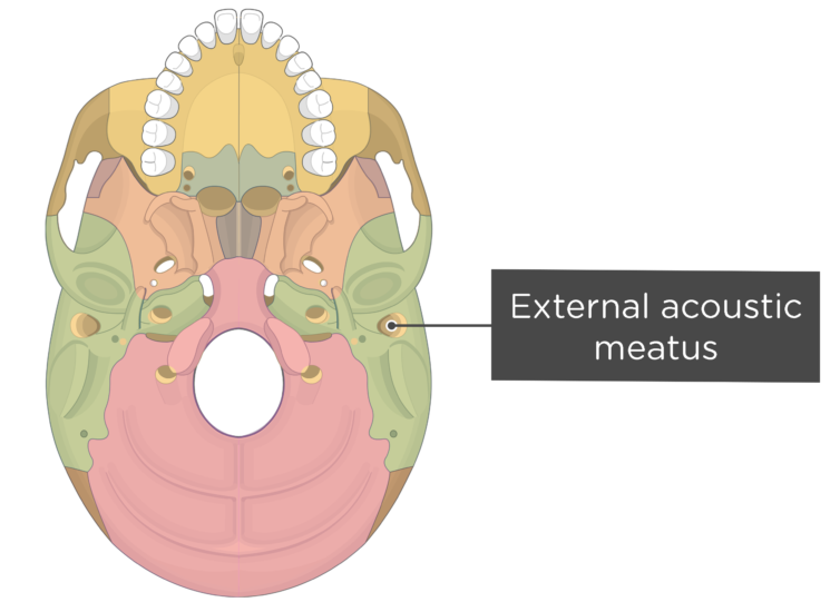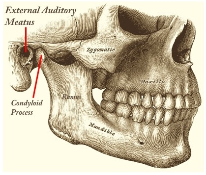
This structure is immediately posterior to the nasopharynx. In every case, the external meatus corresponded to the posterior surface of the clivus, solid bone behind the dorsum sellae, and at the anterior most portion of the basilar occipital bone at its junction with the spenoid bone. The anatomical structure under the mark on the screen was then noted. The consecutive sagittal MRI slices for each individual were examined until the midline slice was found. The middle of the external meatus (as identified by the darkest pixel on the radiographic image within the external meatus image) was marked on the viewing screen for each participant. MRI sagittal slices were taken starting from the external auditory meatus through the midline plane in each of the volunteers. To reduce this, a referencing system using the height of the third cervical vertebrae was used. Positioning on the MRI table and variations in the dimensions of the participants' head and neck will induce errors into any comparisons. T1-weighted images were obtained in the sagittal plane. The acquisition technique was a spin echo sequence with a repetition time of 750 ms and echo time of 11 ms. 4Ī Magnetom Avanto 3T MRI scanner (Siemens Trio, Medical Solutions, Erlangen, Germany) with an anterior flex receiver and posterior coil elements was used.

Two head and neck positions were studied: (i) the neutral head position, obtained by lying on a flat surface (MRI stretcher) without head extension or neck flexion (vertical gaze) and (ii) the sniffing position with neck flexion of 35° and face plane extension of 15° measured using a protractor. The unanaesthetized volunteers underwent magnetic resonance imaging (MRI) of the head and neck region.

a modified Mallampati score of 3 or 4, limited mouth opening, thyromental distance <6 cm, limited neck movement, or upper airway disease), or both were excluded. Participants with a past history or clinical signs of potential difficult direct laryngoscopy, tracheal intubation (i.e. Standard preoperative airway assessment was used to determine anatomic predictors of difficult airway management. Is the ramped position for the obese patient with a higher head position in relation to their antero-posterior chest diameter simply part of this continuum?Īfter local institutional ethics committee approval, informed consent was obtained from 10 adult volunteers ASA grade I–II to participate in a pilot study determining the parts of the airway passage which correspond to the external meatus and sternal notch. The antero-posterior diameter of many obese patients' chests is frequently increased due to large fat deposits across their upper back which leads to a lower head position if one pillow is used for laryngoscopy. Are these secondary markers applicable to non-obese patients? No head elevation is required for neonates to achieve the sniffing position because of their relatively large heads and smaller antero-posterior chest diameters and non-obese adults usually require one standard pillow because of their large head:chest size ratio. This position does produce excellent results in obese patients, but there remains a need to show why these secondary markers are linked to changes in airway configuration with different head and neck positions. The ramped position in this study was defined as horizontal alignment of the external auditory meatus and the sternal notch. The authors did not comment on anatomical changes of the airway apart from the improvement in the laryngoscopy grade. 3 Collins and colleagues stated, ‘It is possible that ramping obese patients produces the same alignment of the axes of intubation that the sniffing position produces in normal weight patients’. 1, 2 The optimal position for laryngoscopy in morbidly obese patients has been described as the ‘ramped position’. Optimal positioning of the head and neck in the sniffing position is governed by the ability to flex the lower cervical spine and extend the occipito-atlanto-axial complex.

Editor-The ‘sniffing position’ has been the cornerstone of direct laryngoscopy and intubation for more than 70 yr.


 0 kommentar(er)
